


COMPARATIVE STUDY OF ANTIBIOTIC RESIDUE, FATTY ACID PROFILE AND MICROBIAL CONTAMINATION IN THE CHEST PORTION OF GALLUS GALLUSDOMESTICUS (BROILER CHICKEN) AND GALLUS GALLUS (TRADITIONAL CHICKEN)
HTML Full TextCOMPARATIVE STUDY OF ANTIBIOTIC RESIDUE, FATTY ACID PROFILE AND MICROBIAL CONTAMINATION IN THE CHEST PORTION OF GALLUS GALLUSDOMESTICUS (BROILER CHICKEN) AND GALLUS GALLUS (TRADITIONAL CHICKEN)
I. Nirmaladevi and K. Prabu *
Department of Biochemistry, Indo-American College, Cheyyar - 604407, Tamil Nadu, India.
ABSTRACT: The modern breeds of chicken (Gallus gallusdomesticus) is supposed to be evolved in India from Red Jungle fowl (G. gallus), whose original habitat is South and Central India, the Himalayan Terrain, Assam, Burma, Ceylon, Sumatra, and Java. Domestication of chicken is believed to have taken place between 7,000 and 10,000 years ago. A part from G. gallus scientists has identified three closely related species that might have bred with the red jungle fowl. It is concluded from this research work; It is well documented that microbial contamination in E. coli, Salmonella, S. aureus respectively and antibiotic residue contamination of chicken with pathogens is a major public health concern worldwide. Based on the Gas chromatography study, traditional chicken has very significant fatty acid profile compared to broiler chicken.
Keywords: Gallus gallusdomesticus, Gallus gallus, Fatty acid profile, Antibiotic residue
INTRODUCTION: Chickens (Gallus gallus-domesticus) are gregarious, omnivorous, ground-dwelling birds that in their natural surroundings search among the leaf litter for seeds, invertebrates, and other small animals. They are one of the most common and widespread domestic animals and is the second most widely eaten type of meat globally. The domestic chicken is descended primarily from the red jungle fowl (Gallus gallus) and is scientifically classified as the same species. As such it can and does freely interbreed with populations of Red jungle fowl 1. The chicken (Gallus gallusdomesticus) is a domesticated fowl, a subspecies of the red jungle fowl.
As one of the most common and widespread domestic animals 2. "Chicken" originally referred to chicks, not the species itself. The species as a whole was then called domestic fowl, or just fowl. Antibiotics are commonly used in the poultry industry for the treatment and prevention of respiratory diseases and other bacterial infections; often administered to groups of poultry via their drinking water.
Poultry and poultry meat are often found contaminated with potentially pathogenic micro-organisms such as Salmonella, Campylobacter, S. aureus, E. coli, and Listeria. The meat surface does not normally, inherently contain pathogenic organisms but can acquire the organisms from fecal matter or from cross-contamination during slaughter. Both poultry muscle and skin are excellent substrates for supporting the growth of a wide variety of microorganisms. Since poultry meat is usually not consumed raw, these outbreaks are caused by undercooking or cross-contamination of ready-to-eat products with microbial contaminants from the raw poultry or others introduced during preparation of the food. Chicken has been used as a suitable model for lipid metabolism studies because dietary modifications, especially dietary fat type can change chicken body composition. Fats act as a condensed source of energy and certain fatty acids such as polyunsaturated fatty acids (PUFAs) are required for both animal and human health.
MATERIALS AND METHODS:
Collection of Sample: A chest portion of broiler and traditional chickens were collected from local retail shops in Cheyyar. The collected samples were kept in separate plastic bags, transferred directly to the laboratory under the aseptic conditions.
Preparation of Samples: Twenty-five grams of the examined samples were removed by sterile scissors and knife. Each sample was cut into small fine spices and homogenized the samples by using motor and pestle. The homogenate samples were centrifuged at 2000 rpm for 1-3 min. Finally, collect both samples.
Antibiotic Detection: The Kirby Bauer method (Antibiotic) was used. The MHA plates were used. Three species of bacteria were used: Escherichia coli, Staphylococcus aureus, and Pseudomonas sp.
Microbial Contamination:
Serial Dilution: Homogenized samples were taken in the 1 ml of was transferred with a sterile pipette to a sterile test tube containing 9 ml of sterile peptone water and mixed well to make the next dilution. This procedure was repeated and using a sterile pipette for each dilution up to 10-9 dilution. After using the spread plate method.
Biochemical Test: The bacterial isolates were identified based on the biochemical tests. The IMVIC method used it.
Determination of Fatty Acids Profile:
Gas Chromatography: An Agilent 6890 gas chromatograph was equipped with a straight deactivated 2 mm direct injector liner and a 15m Alltech EC-5 column (250μ I.D., 0.25μ film thickness). A split injection was used for sample introduction, and the split ratio was set to 10:1. The oven temperature program was programmed to start at 35 ºC, hold for 2 min, then ramp at 20 ºC per min to 300 ºC and hold for 5 min. The helium carrier gas was set to 2 ml/min flow rate (constant flow mode).
Mass Spectrometry: A JEOL GCmate II benchtop double-focusing magnetic sector mass spectrometer operating in electron ionization (EI) mode with TSS-20001 software was used for all analyses. Low-resolution mass spectra were acquired at a resolving power of 1000 (20% height definition) and scanning from m/z 25 to m/z 700 at 0.3 sec per scan with a 0.2-second inter-scan delay. High-resolution mass spectra were acquired at a resolving power of 5000 (20% height definition) and scanning the magnet from m/z 65 to m/z 750 at 1 sec per scan.
Mass Spectrometry Library Search: Identification of the components of the purified compound was matching their recorded spectra with the data bank mass spectra of NIST library V 11 provided by the instrument's software.
Quantitative Analysis:
- Estimation of Glucose by Ortho-Toluidine Method.
- Estimation of Protein by Lowry’s Method.
RESULTS:
Detection of Antibiotic Residues: Screening of Antibiotic Residue in Broiler Chicken and Traditional Chicken is studied on determine presence the zone in diameter.
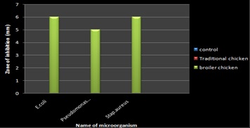
FIG. 1: ANTIBIOTIC RESIDUE DETECTION OF CHEST PORTION IN GALLUS GALLUS AND GALLUS GALLUSDOMESTICUS
The zone indicates the presence of antibiotic residue. This method used three different microorganisms like E. coli, Pseudomonas, and S. aureus. However, as a result in Table 1 and Fig. 1. The zoning measure at mm.
Study of Microbial Contamination: Determination of microbial contamination in both broiler chicken and traditional chicken in indicating the presence of colonies. And further used in the biochemical test for IMVIC method. Show on Fig. 2, 3 and 4 respective for microbial contamination and biochemical test.
Biochemical Analysis: Quantitative Analysis of Glucose and Protein in Broiler chicken Traditional chicken. The results are shown in Table 2 and Fig. 5.
TABLE 1: ANTIBIOTIC RESIDUE DETECTION OF CHEST PORTION IN GALLUS GALLUS AND GALLUS GALLUSDOMESTICUS
| Zone of Inhibition in Diameter | |||
| Microorganism
name |
Control | Gallus gallus
(Traditional chicken sample) |
Gallus gallusdomesticus (Broiler chicken sample) |
| E. coli | 0 mm | 0 mm | 6 mm |
| S. aureus | 0 mm | 0mm | 6 mm |
| Pseudomonas sps | 0 mm | 0 mm | 5 mm |

FIG. 2: MICROBIAL CONTAMINATION OF CHEST PORTION IN (A) GALLUS GALLUS AND (B) GALLUS GALLUSDOMESTICUS
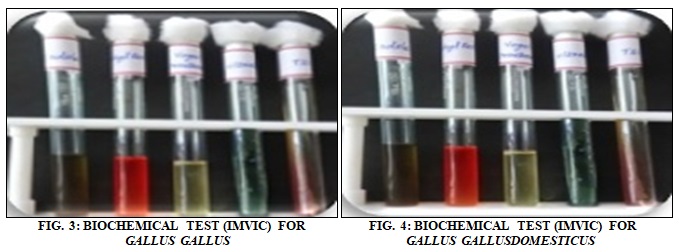
TABLE 2: QUANTITATIVE ANALYSIS OF BIOCHEMICALS IN GALLUS GALLUS AND GALLUS GALLUSDOMESTICUS
| S.
no. |
Substances | Gallus gallus
(broiler chicken) |
Gallus gallusdomesticus
(traditional chicken) |
| Chest portion (mg %) | Chest portion (mg %) | ||
| 1 | Total glucose | 5.937 | 0.156 |
| 2 | Total proteins | 12 | 9 |
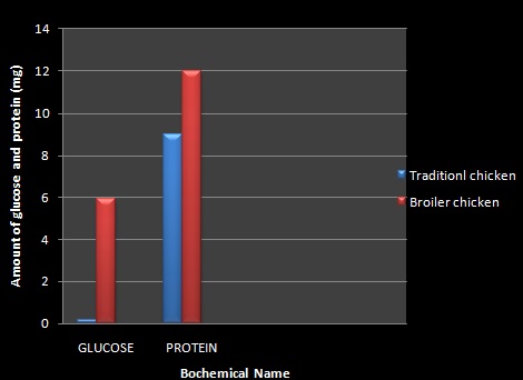
FIG. 5: QUANTITATIVE ANALYSIS OF BIOCHEMICAL IN THE CHEST PORTION OF GALLUS GALLUS AND GALLUS GALLUSDOMESTICUS
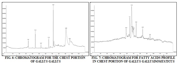
Determination of Fatty Acids: Screening of Fatty Acids in Both chickens for Gas chromatography methods. However, results shown in Table 3, 4 respective for broiler chicken, traditional chicken, and Fig. 6, 7 respective for chromatogram in broiler chicken and traditional chicken.
TABLE 3: DETERMINATION OF FATTY ACIDS IN THE CHEST PORTION OF GALLUS GALLUS
| Peak no. | RT (min) | Compound name | Peak area | Peak area (%) |
| 1 | 9.42 | Tetradecanoic acid | 18051088 | 8.82 |
| 2 | 12.12 | 10-Octadecenal | 31631552 | 15.45 |
| 3 | 14.27 | 1-Dodecanol,3,7,11-trimethyl- | 12804160 | 6.25 |
| 4 | 17.23 | Pentadecanoic acid, 14-methyl-, methyl ester | 21011008 | 10.26 |
| 5 | 18.97 | n-Hexadecanoic acid | 67618784 | 33.02 |
| 6 | 23.85 | 16-Octadecenoic acid, methyl ester | 34268256 | 16.74 |
| 7 | 25.22 | 9-Octadecenoic acid[Z]-, hexyl ester | 19374064 | 9.46 |
| Total | 204758912 | 100.00 |
TABLE 4: DETERMINATION OF FATTY ACIDS IN THE CHEST PORTION OF GALLUS GALLUSDOMESTICUS
| Peak no. | RT (min) | Compound name | Peak area | Peak area (%) |
| 1 | 15.75 | Tetradecanioc acid | 18464256 | 10.57 |
| 2 | 17.1 | Pentadecanoic acid, 14- methyl- methyl ester | 26749136 | 15.31 |
| 3 | 17.83 | Icosadienoic acid | 35950272 | 20.58 |
| 4 | 18.43 | n-Hexadecanoic acid | 19329488 | 11.07 |
| 5 | 18.48 | Estra-1,3,5 [10]-trien-17 a-o1, | 18377024 | 10.52 |
| 6 | 19.53 | 10-Octadecenol, | 22790896 | 13.05 |
| 7 | 22.02 | 9-Octadecenoic acid[Z]-, hydroxyethyl ester | 19747376 | 11.30 |
| 8 | 24.8 | Docashexaenoic acid[Z]-hexyl ester | 13273264 | 7.64 |
| Total | 174681712 | 100.00 |
DISCUSSION AND CONCLUSION: These resistant strains colonize the human intestine, and the genes coding resistance to antibiotics can be transferred to bacterial strains that belong to natural microflora 3. Applications of antibiotics bring about an increase in resistance to antibiotics not only in pathogenic bacterial strains but also in strains forming a part of the endogenous flora of humans and animals4. And also confirmed that in chickens fed with tetracycline there was a transfer of tetracycline resistance genes between chicken Escherichia coli strains, from chicken to chicken and from chicken to man 5.
Antibiotic usage in farm animals has raised many concerns, among which the potential transfer of antibiotic-resistant pathogens from animals to humans 6. This transfer has severe health implications, including treatment failures, which has led to some deaths and increased the cost of human therapies 7. Furthermore, overuse of antibiotics leads to the occurrence of harmful residues in edible poultry tissues (meat and eggs) and other animal products 6. The global human community has an ongoing and worsening crisis of antibiotic-resistant infections in patients.
It is important that we not be bogged down or distracted by quibbles over the minutiae of the molecular mechanisms by which antibiotic resistance spreads from animals to humans or the precise proportion of antibiotic-resistant infections in humans that is caused by antibiotic use in animals. The most important cause of antibiotic resistance is the consumption of antibiotic as a growth promoter and treatment for animals and human. Therefore, it is very important to decrease the consumption of antibiotic in animal and humans.
It is well documented that contamination of food with pathogens is a major public health concern worldwide. Fresh markets are traditional open-air markets where chickens are sold by individual vendors or farmers, and often sold and stored at ambient temperatures. These markets naturally have multiple sources of potential contamination (rodents, insects, sewage). Raw poultry meats are commonly contaminated with E. coli; this is particularly true of chicken products. Broilers arriving at the poultry slaughterhouse for processing are generally highly contaminated with bacteria, especially with potential human pathogenic bacteria, such as Coliform and Salmonella 8. Microbial contamination of poultry carcasses is a natural result of different procedures necessary to produce retailed products from living birds. Most of the bacterial contaminants are nonpathogenic; however, poultry is known to harbor a large number of bacteria that are pathogenic to human being 9. Contamination takes place during the handling and preparation of the meat and also from air dust and personal contact, external sources during bleeding, handling, and cutting. Additional contamination took place in the retails markets and containers 10. The broiler and traditional chicken samples sold for human consumption were screened for antibiotic residues, and results are illustrated in Table 2. The zone size of antibiotic positive chicken samples measured in diameters is presented in Fig. 4, 5, 6.
Drug residues in foods are of a major public health concern in many countries, especially where most food sales bypass official quality assurance channels. Consumers are very much conscious that their food supply is free of contamination by herbicides, pesticides, drugs, and antibiotics because they may cause severe health hazards, causing allergic reactions, carcinogenicity and promotion of the spread of bacterial resistance to antibiotics used in human medicines. Real and perceived concerns about the harmful consequences of drug residues in food have created an important need for monitoring the food. Monitoring of antibiotic residues in animal-derived foods is essential to protect human health. Therefore the present approach for monitoring of antibacterial drugs has been hypothesized, and the samples of chicken meat were collected to detect antibiotic residues.
The residual contamination appeared high in the broiler chicken. This might indicate that there may be prevalent use of antibacterial drugs in chicken animals and after treatments; withdrawal period of the drug was not properly followed in these areas. In consequence of such results Sasanya et al., reported that ninety percent (90%, 105/117) of the respondents acknowledged the use of antibiotics on their animals but ninety-six percent (96%, 112/117) of the respondents did not observe drug withdrawal periods in animals or animal products sold from their farms, while only 14% (16/117) of the respondents were aware of the human health risks associated with exposure to residues of drugs through consumption of contaminated animal products. In the present study compared to tradition chicken chest portion of the broiler chicken were observed as more contaminated with the antibiotic residues. Similarly, Ibrahim et al. reported that over 50 slaughtered cattle at Sokoto metropolitan abattoir 44% of the slaughtered cattle were detected positive to antibiotic residues using E. coli, Staphylococcus aureus, and Pseudomonas test.
In another study 43% of meat samples were reported to be contaminated with sulfonamide residues; However, in current study the high level of contamination noted in broiler chicken meat samples could be due to the massive use, uncontrolled and prolonged antibiotics at livestock farms by farmers with or without guidance of veterinarians to prevent or treat the diseases. Such residual contamination also might be due to the early slaughtering of animals after administration of drugs and withdrawal periods of drugs are not followed. The chromatogram Fig. 6 revealed the presence of FA’s namely Tetradecanoic acid, 10-Octadecenal,1-Dodecanol,3,7,11-trimethyl-, Penta-decanoic acid, 14-methyl-, methyl ester, n-Hexadecanoic acid, 16-Octadecenoic acid, methyl ester, 9-Octadecenoic acid[Z]-, hexyl ester were present in the Traditional chicken. A higher level of (33.02%) n-Hexadecanoic acid and (15.45%) 10-Octadecenal was observed in the chest tissues of traditional chicken.
The chromatogram Fig. 7 revealed the presence of FA’s namely Tetradecanioc acid, Pentadecanoic acid, 14- methyl- methyl ester, Icosadienoic acid, n-Hexadecanoic acid, Estra-1,3,5 [10]-trien-17 a-o1, 10-Octadecenol, 9-Octadecenoic acid[Z]-, hydroxyethyl ester, Docashexaenoic acid[Z]-hexyl ester were present in the Broiler chicken. A higher level of (20.58%) Icosadienoic acid and (15.31%) Pentadecanoic acid, 14- methyl- methyl ester was observed in the chest tissues of broiler chicken. The protein, glucose, content of Traditional chicken were analyzed in chest tissues in the following study were as follows: Protein (9 mg), glucose (0.156 mg) respectively. The protein, glucose, content of Broiler chicken were analyzed in chest tissues in the following study were as follows: Protein (12 mg), glucose (5.937 mg) respectively
ACKNOWLEDGEMENT: Nil
CONFLICT OF INTEREST: Nil
REFERENCES:
- Wong GK: A genetic variation map for chicken with 2.8 million single-nucleotide polymorphisms. (https:// www.ncbi.nlm.nih.gov/pmc/articles/PMC226312) 2004.
- Eduard W: Exposure to non-infectious microorganisms and endotoxins in agriculture. Ann Agric Environ Med 1997; 4: 179.
- Bogaard AE and Stobberingh EE: Epidemiology of resistance to antibiotics. Links between animals and humans. Int J Antimicrob Agents 2000; 14: 327-335.
- Kolar M: Occurrence of antibiotic-resistant bacteria strains isolated in poultry- (2002) M.V.Sc. Thesis University of Khartoum, the Sudan 2004.
- Levey SB, Fitzgerald GB and Macone AB: Spread of antibiotic resistance plasmids from chicken to chicken and from chicken to man. Nature 1976; 260: 400-421.
- Oluwawemimo OA Antibiotic use and practices in commercial poultry laying hens in Ogun state Nigeria 2016; 72: 1.
- Levey SB, Fitzgerald GB and Macone AB: Spread of antibiotic resistance plasmids from chicken to chicken and from chicken to man. Nature 1976; 260: 400-421.
- Göksoy EÖ, Kirkan S and Kök F: Microbiological quality of broiler carcasses during processing in two slaughterhouses in Turkey. Poultry Science 2004; 83(8): 1427-1432.
- Zhang L, Davis MA and Conner DE: Poultry-borne pathogens plant 2001.
- Harrigan WF and McCance ME: Laboratory Methods in Food and Dairy Microbiology. Academic Press, London 1976.
Nirmaladevi I and Prabu K: Comparative study of antibiotic residue, fatty acid profile and microbial contamination in the chest portion of Gallus gallusdomesticus (Broiler chicken) and Gallus gallus (Traditional chicken). Int J Life Sci & Rev 2018; 4(7): 104-09. doi: 10.13040/ IJPSR.0975-8232.IJLSR.4(7).104-09.
All © 2015 are reserved by International Journal of Life Sciences and Review. This Journal licensed under a Creative Commons Attribution-NonCommercial-ShareAlike 3.0 Unported License.