


THE EFFECT OF GLUCOSE ON GROWTH OF MESENCHYMAL STEM CELLS DERIVED FROM UMBILICAL CORDS OF NORMAL AND GESTATIONAL DIABETIC MOTHERS.
HTML Full TextTHE EFFECT OF GLUCOSE ON GROWTH OF MESENCHYMAL STEM CELLS DERIVED FROM UMBILICAL CORDS OF NORMAL AND GESTATIONAL DIABETIC MOTHERS
Nadia Wajid * 1, Noreen Latief 2, Muhammad Ali 1, Sara Javed 1, Rashida Naseem 1 and Fatima Ali 1
Institute of Molecular Biology and Biotechnology 1, The University of Lahore, Lahore, Pakistan.
CEMB 2, University of the Punjab, Lahore, Pakistan.
ABSTRACT: Objective: To explore the effects of glucose concentration on the growth of Wharton’s jelly mesenchymal stem cells from umbilical cords of gestational diabetic mothers (DWJMSCs) and normal mothers (NWJMSCs). Methods: NWJMSCs and DWJMSCs were treated at passage 3 with low (5.5mM) or high glucose. Cells’ growth was evaluated by methyl thiazolyl tetrazolium uptake and cytotoxicity by LDH release. Status of oxidative stress was estimated by the release of stress enzymes, i.e., glutathione s transferase (GSH) and catalase. Results: The results of cells’ growth assays indicated a significant increase in proliferation in DWJMSCs when treated with high glucose as compared to low glucose while the opposite was found in NWJMSCs. Conversely, cytotoxicity was lower in DWJMSCs treated with 25mM glucose while it was increased in NWJMSCs at the same concentration and vice-versa. Both groups of cells exhibited similar behavior in the release of antioxidant enzymes, i.e., GSH reduced in both cell types at 25mM glucose while no difference was observed in catalase activity. Conclusion: Growth of DWJMSCs is enhanced at high glucose concentrations while it is reduced in NWJMSCs.
Keywords: MSCs, Umbilical cord, Gestational diabetes, High glucose, Low glucose
INTRODUCTION: Stem cells are essential tools for tissue engineering for the repair of various biological structures including skin, smooth muscle tissue, blood vessels, cardiac tissue, renal tubules, intestine, bladder subunits, bones, etc. as they can differentiate into multiple cell types 1. Human umbilical cords derived Wharton’s jelly is a rich source of mesenchymal stem cells which are (WJMSCs) are well-tolerated by immune system 2, 3.
In-vitro expansion, which is inevitable for therapeutics is limited because of in-vitro aging due to the induction of stress by culture conditions leads to replicative senescence. So culture conditions must be optimized to avoid such conditions and improve the proliferation of isolated cells 4. Glucose is not only an essential source of energy for cells but also acts as a substrate for the synthesis of protein and lipid.
Many pathological conditions, including diabetes mellitus, are associated with hyperglycemia 5 and oxidative stress, which affects the growth of stem cell 6. It is reported that WJMSCs derived from diabetic mothers (DWJMSCs) show a reduced growth rate in culture conditions as compared to normal mothers’ derived WJMSCs (NWJMSCs) 7. This study is based on finding the optimal glucose concentration for maximum growth of NWJMSCs and DWJMSCs in-vitro.
MATERIALS AND METHODS:
Procurement of Human Umbilical Cord: The study was approved by the biosafety board at The University of Lahore, Lahore, Pakistan. Mothers selected for the study were negative for human immunodeficiency virus (HIV), hepatitis B and C virus (HBV and HCV). Umbilical Cords were obtained with the consent of the parents from full-term caesarian sections.
Isolation, Culturing, and Characterization of WJMSCs: WJMSCs were isolated by explant culturing method from the human umbilical cord of mothers with or without gestational diabetes as previously described by us 7. Briefly, 5 g cord tissue was cut in to smaller pieces which were subsequently incubated in Dulbecco’s modified eagle medium-low glucose (DMEM LG)(Sigma Aldrich, USA) with 10 % fetal bovine serum(FBS) (Gibco, Grand Island, NJ) and 100 U/ml penicillin and 100 µg/ml streptomycin (Gibco, Grand Island, NJ). Culture medium was renewed after every three days. At passage three DWJMSCs were plated at a density of 100,000 cells per well of a6-well plate (Corning, USA). After 3 days of culturing cells of both types were divided into two groups, i.e. provided with high glucose DMEM (25mM) (Sigma Aldrich, USA) or provided with low glucose DMEM (5.5mM) (Sigma Aldrich, USA). The culture medium after day 3 was saved at -80 ºC for LDH assay and oxidative stress while cells were used for MTT assay.
Cell Proliferation Assay: To compare the proliferative potential of DWJMSCs and NWJMSCs in both groups, the 3-(4, 5-dimethyl thiazol-2-yl)-2, 5-diphenyltetrazolium bromide (MTT) assay was performed. A monolayer of cells was first washed with phosphate buffer saline (PBS) (In vitro gen Inc., USA). 500 µL complete medium along with 60 µL MTT solution (In vitro gen Inc., USA) was added to cells and incubated for 2 hours at 37°C. Purple color crystals formed within cells were solubilized with DMSO and absorbance was taken at 570 nm.
LDH Assay: LDH assay was performed using 5µL medium from each group at the end of treatment using LDH assay kit (AMP Diagnostics, Austria) according to the manufacturer’s instructions. Briefly, 5µL cell culture medium of both groups was mixed with 95 µL working reagent, incubated for 5 min and then absorbance was recorded at a wavelength of 340 nm.
Estimation of Glutathione: Amount of reduced glutathione (GSH) in culture medium after treatment was estimated using the method of 7, 8. For this, 0.5 mL culture medium was added in a tube along with 2.0 mL disodium hydrogen phosphate buffer (0.3 M) and 0.25 mL 5,5'-dithiobis-(2-nitrobenzoic acid) or DTNB (0.001 M) (In-vitro gen Inc., USA). The mixture was incubated for 15 min, and absorbance was measured at 412 nm.
Estimation of Catalase Activity: The activity of catalase was monitored by using the method described by Sinha 9. 0.1 mL culture medium was taken and mixed with 1.0 mL phosphate buffer (10 mM, pH 7.0) and 0.4 mL H2O2 (0.2 M) (Sigma Aldrich, USA). The reaction was stopped by adding 2.0 mL dichromate acetic acid reagent. Samples were incubated for 10 min in a boiling water bath, cooled and absorbance was measured at 530 nm.
Statistical Analysis: Three independent experiments in triplicates were conducted for obtaining quantitative data. Statistical analysis was performed using Graph Pad Prism version 5.00 for Windows (Graph Pad Software, San Diego California USA). All results were expressed as mean ± standard deviation (SD). Student’s t-test was used for comparison. Statistical significance was considered as P<0.05.
RESULTS:
Effect of Glucose Concentration on Cells’ Proliferation: Cells’ proliferation was assessed by MTT assay. It was observed that DWJMSCs cultured in LG DMEM (group 2) showed a significantly lower proliferation compared to DWMSCs cultured in HG DMEM while the opposite was observed in NWJMSCs Fig. 1 & 2.
Cytotoxicity Evaluation: Cytotoxicity was analyzed by LDH release. It was observed that in NWJMSCs LDH release was higher at 25mM glucose concentration as compared to low glucose i.e. 5.5mM.
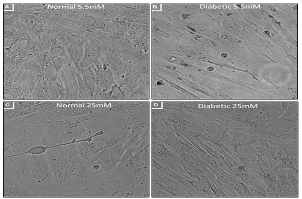
FIG. 1: PHASE CONTRAST MICROSCOPY OF NWJMSCS AND DWJMSCS CULTURED IN LOW AND HIGH GLUCOSE. NWJMSCs APPEAR HEALTHY IN 5.5mm GLUCOSE WHILE DWJMSCS IN 25mm GLUCOSE
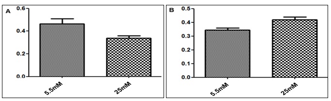
FIG. 2: CELLS’ PROLIFERATION ASSAY. NWJMSCS EXPRESS HIGHER PROLIFERATION IN 5.5mM GLUCOSE COMPARED TO 25mM GLUCOSE WHILE OPPOSITE WAS OBSERVED IN DWJMSCS
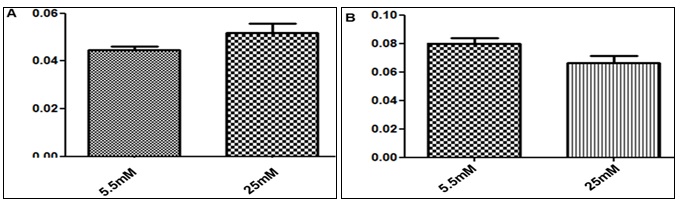
FIG. 3: LDH ASSAY. NWJMSCs EXPRESS LOWER DEATH RATE IN 5.5mM GLUCOSEWHILE DWJMSCs IN 25mM
In contrast in DWJMSCs LDH release was significantly lower at 25mM glucose Fig. 3. Oxidative Stress: Oxidative stress was evaluated in NWJMSCs and DWJMSCs treated with high and low glucose. It was observed that there was no significant difference between antioxidant enzymes, i.e. level of GSH was reduced at 25mM, and catalase activities were the same in both study groups Fig. 4.
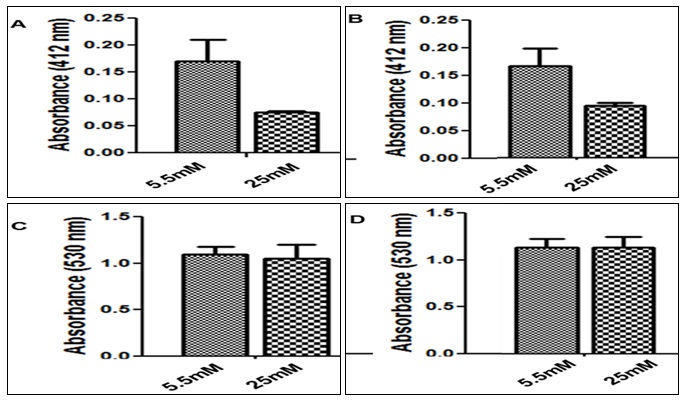
FIG. 4: OXIDATIVE STRESS. (A) GSH ACTIVITY WAS REDUCED IN HIGH GLUCOSEBOTH IN NWJMSCs AND DWJMSCs. (B) THERE WAS NO SIGNIFICANT DIFFERENCE BETWEEN NWJMSCs AND DWJMSCs IN CATALASE ACTIVITY IN HIGH OR LOW GLUCOSE IN CULTURE MEDIUM
DISCUSSION AND CONCLUSION: WJMSCs are considered the ideal source for autologous and allogeneic transplantation and have gained popularity for private and public banking due to high isolation rates, minimum ethical concerns, low immunogenicity and most importantly the postnatal isolation 10. Gestational diabetes mellitus (GDM) impairs the proliferative capacity of WJMSCs. The key factor involved in GDM is an excessive amount of glucose in mother, which crosses freely to fetus 7. To achieve maximum therapeutic benefits, it is a prerequisite to optimizing the culture conditions for cellular growth.
Hence the present study is based on an evaluation of glucose concentration for optimal growth of DWJMSCs in vitro for therapeutic usage. It is necessary to find the cells’ proliferation rate for their clinical usage and reported that high glucose concentration in the culture medium of cells does not affect Proliferation of human mesenchymal stem cells acutely while it has detrimental effects on many cell types 11. We evaluated the cells’ proliferation by MTT assay, which MTT is a positively charged tetrazolium salt and is converted into a purple formazan product after entry into actively metabolizing viable cells 12. It was found that DWJMSCs show an increased growth after treatment with high glucose in culture medium as compared to treatment with low glucose and the opposite was observed in NWJMSCs Fig. 1 & 2. Cells’ death rate is an important tool to analyze the growth rates of a healthy growing population for transplantation, which is measured in routine by LDH assay. Cell death causes the release of this soluble cytosolic enzyme into culture medium 13. We found that DWJMSCs release less LDH when grown in high glucose concentrations while NWJMSCs release more Fig. 3.
Hyperglycemia may promote oxidative stress hence damage to multiple organs in the body, i.e. eyes, kidneys, nerves, and blood vessels. The oxidative stress in body due to high glucose level could be detrimental for stem cells therapy. But it is also reported that high glucose may increase the proliferation of stem cells 5. In our study no significant difference in the release of oxidative stress enzymes (GSH and catalase) was found in NWJMSCs and DWJMSCs, i.e. GSH was reduced significantly while no effect on catalase activity was observed Fig. 3. In conclusion, WJMSCs, when derived from GDM mothers, show increased proliferation, reduced cells ‘death in high glucose culturing condition while they show no significant change in the release of antioxidant stress enzymes while NWJMSCs behave oppositely in same glucose concentrations.
ACKNOWLEDGEMENT: This work was supported by research grants from The University of Lahore, Lahore, Pakistan.
CONFLICT OF INTEREST: The authors declare no conflicts of interest.
REFERENCES:
- Deorosan B and Nauman EA: The role of glucose, serum, and three-dimensional cell culture on the metabolism of bone marrow-derived mesenchymal stem cells. Stem Cells Int 2011; 429187.
- Ma L, Zhou Z, Zhang D, Yang S, Wang J, Xue F, Yang Y, and Yang R: Immunosuppressive function of mesenchymal stem cells from human umbilical cord matrix in immune thrombocytopenia patients. Thromb Haemost 2012: 107(5): 937-950.
- Troyer DL and Weiss ML: Concise review: Wharton’s jelly-derived cells are a primitive stromal cell population. Stem Cells 2008; 26(3): 591-599.
- Stolzing A, Coleman N and Scutt A: Glucose-induced replicative senescence in mesenchymal stem cells. Rejuvenation Res 2006; 9(1): 31-5.
- Saki N, Jalalifar MA, Soleimani M, Hajizamani S and Rahim F: Adverse effect of high glucose concentration on stem cell therapy. Int J Hematol Oncol Stem Cell Res 2013; 7(3): 34-40.
- Khan M, Akhtar S, Mohsin S, Khan N and Riazuddin S: Growth factor preconditioning increases the function of diabetes-impaired mesenchymal stem cells. Stem Cells Dev 2011; 20(1): 67-75.
- Wajid N, Naseem R, Anwar SS, Awan SJ, Ali M, Javed S and Ali F: The effect of gestational diabetes on proliferation capacity and viability of human umbilical cord-derived stromal cells. Cell Tissue Bank 2014; PMID: 25407535.
- Beutler E, Duron O and Kelly BM: Improved method for the determination of blood glutathione. J Lab Clin Med 1963; 61: 882-888.
- Sinha AK: Colorimetric assay of catalase. Anal Biochem 1972; 47: 38994.
- Kalaszczynska I and Ferdyn K: Wharton's jelly-derived mesenchymal stem cells: future of regenerative medicine? Recent findings and clinical significance. Biomed Res Int 2015; 430847.
- Weil BR, Abarbanell AM, Herrmann JL, Wang Y and Meldrum DR: High glucose concentration in the cell culture medium does not acutely affect human mesenchymal stem cell growth factor production or proliferation. Am J Physiol Regul Integr Comp Physiol 2009; 296(6): R1735–R1743.
- Valcheva-Kuzmanova SV, Beronova AB and Momekov GT: Protective effect of Aronia melanocarpa fruit juice in a model of cisplatin-induced cytotoxicity in-vitro. Folia Med (Plovdiv) 2013; 55(3–4): 76–79.
- Chan FK, Moriwaki K and De Rosa MJ: Detection of necrosis by the release of lactate dehydrogenase activity. Methods Mol Biol 2013; 979: 65–70.
How to cite this article:
Wajid N, Latief N, Ali M, Javed S, Naseem R and Ali F: The effect of glucose on growth of mesenchymal stem cells derived from umbilical cords of normal and gestational diabetic mothers. Int J Life Sci & Rev 2015; 1(6): 222-26. doi: 10.13040/IJPSR.0975-8232.IJLSR.1(6).222-26.
All © 2015 are reserved by International Journal of Life Sciences and Review. This Journal licensed under a Creative Commons Attribution-NonCommercial-ShareAlike 3.0 Unported License.
Article Information
4
222-226
856
2011
English
IJLSR
N. Wajid*, N. Latief, M. Ali, S. Javed, R. Naseem and F. Ali
Institute of Molecular Biology and Biotechnology, The University of Lahore, Lahore, Pakistan
Nadia.wajid@imbb.uol.edu.pk
10 May 2015
24 June 2015
28 June 2015
10.13040/IJPSR.0975-8232.IJLSR.1(6).222-26
30 June 2015